45 microscope diagram without labels
Labelled Diagram of Compound Microscope - Biology Discussion The below mentioned article provides a labelled diagram of compound microscope. Part # 1. The Stand: The stand is made up of a heavy foot which carries a curved inclinable limb or arm bearing the body tube. The foot is generally horse shoe-shaped structure (Fig. 2) which rests on table top or any other surface on which the microscope in kept. Compound Microscope Parts - Labeled Diagram and their Functions - Rs ... The eyepiece (or ocular lens) is the lens part at the top of a microscope that the viewer looks through. The standard eyepiece has a magnification of 10x. You may exchange with an optional eyepiece ranging from 5x - 30x. [In this figure] The structure inside an eyepiece. The current design of the eyepiece is no longer a single convex lens.
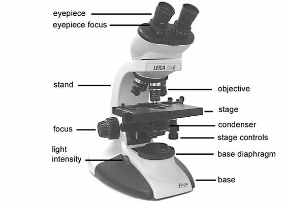
Parts of a microscope with functions and labeled diagram Figure: Diagram of parts of a microscope There are three structural parts of the microscope i.e. head, base, and arm. Head - This is also known as the body. It carries the optical parts in the upper part of the microscope. Base - It acts as microscopes support. It also carries microscopic illuminators.

Microscope diagram without labels
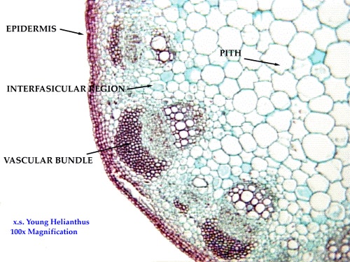
Amazing 27 Things Under The Microscope With Diagrams Amazing 27 Things Under The Microscope With Diagrams February 20, 2022 by Anupama Sapkota Note: Each image source is given below in this post of respective subheadings. Table of Contents 1. Amoeba under the microscope Direct observation Observation after staining 2. Algae under the microscope Chlorophyta Chromophyta Cryptophyta Rhodophyta How Does a Microscope Work A compound microscope has two or more lenses. The eyepiece or ocular lens sits atop the body tube. Many microscopes are binocular and have two ocular lenses. Additionally, a binocular head will have a prism, either in the head or the body tube, to split the image and direct it to both oculars. Junqueira's Basic Histology Text and Atlas, 14th Edition Enter the email address you signed up with and we'll email you a reset link.
Microscope diagram without labels. Label Microscope Diagram - EnchantedLearning.com Using the terms listed below, label the microscope diagram. arm - this attaches the eyepiece and body tube to the base. base - this supports the microscope. body tube - the tube that supports the eyepiece. coarse focus adjustment - a knob that makes large adjustments to the focus. diaphragm - an adjustable opening under the stage, allowing ... Microscope Labeling - The Biology Corner Students label the parts of the microscope in this photo of a basic laboratory light microscope. Can be used for practice or as a quiz. ... The type of microscope used in most science classes is the _____ microscope. 18. You should carry the microscope by the _____ and the _____. 19. The objectives are attached to what part of the microscope ... Microscope Labeling Game - PurposeGames.com About this Quiz. This is an online quiz called Microscope Labeling Game. There is a printable worksheet available for download here so you can take the quiz with pen and paper. This quiz has tags. Click on the tags below to find other quizzes on the same subject. Science. Label the microscope — Science Learning Hub In this interactive, you can label the different parts of a microscope. Use this with the Microscope parts activity to help students identify and label the main parts of a microscope and then describe their functions. Drag and drop the text labels onto the microscope diagram.
Microscope Parts and Functions First, the purpose of a microscope is to magnify a small object or to magnify the fine details of a larger object in order to examine minute specimens that cannot be seen by the naked eye. Here are the important compound microscope parts... Eyepiece: The lens the viewer looks through to see the specimen. Microscope, Microscope Parts, Labeled Diagram, and Functions The Iris Diaphragm is located above the condenser lens and below the microscope stage. The different sized holes in the diaphragm helps to vary the size of the cone and intensity of light that is projected upward into the slide. However, there is no set rule regarding which setting to use for a particular power. Parts of the Microscope with Labeling (also Free Printouts) Parts of the Microscope with Labeling (also Free Printouts) A microscope is one of the invaluable tools in the laboratory setting. It is used to observe things that cannot be seen by the naked eye. Table of Contents 1. Eyepiece 2. Body tube/Head 3. Turret/Nose piece 4. Objective lenses 5. Knobs (fine and coarse) 6. Stage and stage clips 7. Aperture Microscope With Labels clip art - clker.com Download Clker's Microscope With Labels clip art and related images now. Multiple sizes and related images are all free on Clker.com. Facebook Login; X. E-mail Password. ... microscope diagram blank; microscope drawing without label; microscope and its label; symbol of microscope; blank microscope to label; compound microscope side view;
7th grade Science - Microscope Diagram - Quizlet The Parts of a Microscope. 12 terms. totobear PLUS. Sets found in the same folder. Science Key terms 7th grade. 13 terms. palocastillo. 7th Grade Earth Science. 9 terms. EliseC17. 7thGrade Review - Cells/Biology. 26 terms. SolizScience TEACHER. 7th grade Science, Cell theory. 8 terms. Super1412. Other sets by this creator. Worksheet Student - Worksheet Website for Students May 01, 2022 · Worksheet Website for Students. The worksheets include first grade appropriate reading passages and related questions. 1st grade reading comprehension worksheets printable pdf today the number of words that can... Microscope Foldable Worksheets & Teaching Resources | TpT Microscope Mini-Bundle - Foldable, Powerpoint, and 2 Lab Activities. by. The Skye World Science. 1. $9.00. $6.00. Bundle. Zip. This mini-bundle of microscope activities will prepare your 7th grade science and 10th grade biology students to identify the parts and functions of a microscope, view objects properly, and set up wet mount slides. Label the Microscope Diagram | Download Scientific Diagram In the study of antibiogram, all isolates were shown 100% sensitive to streptomycin, ciprofloxacin and chloramphenicol. Maximum 41% isolates were shown resistant to co-trimethaxozole whereas 30% ...
33 Microscope Diagram To Label - Labels Database 2020
Labeling Microscope Worksheet | Teaching Resources A straightforward worksheet in which students are required to identify the parts of a basic microscope. Tes classic free licence. Reviews. 4.7 Something went wrong, please try again later. MACS0647-JD. a year ago. report. 5. Thanks. Very helpful. Empty reply does not make any sense for the end user ...
The Microscope - General Revision for GCSE
Animal Cell Simple Labeled Diagram - Mitosis Diagram Without Labels For ... Cells communicate with one another and are responsible for transmitting microscope label the diagram of a microscope. A comparison of plant and animal cells using labelled diagrams and descriptive explanations. ... Mitosis diagram without labels for kids simple animal cell. The animal cell diagram is widely asked in class 10 and 12 examinations ...
8 Best Images of Lens Diagram Worksheet - Microscope with Labeled Parts, Label Eye Parts ...
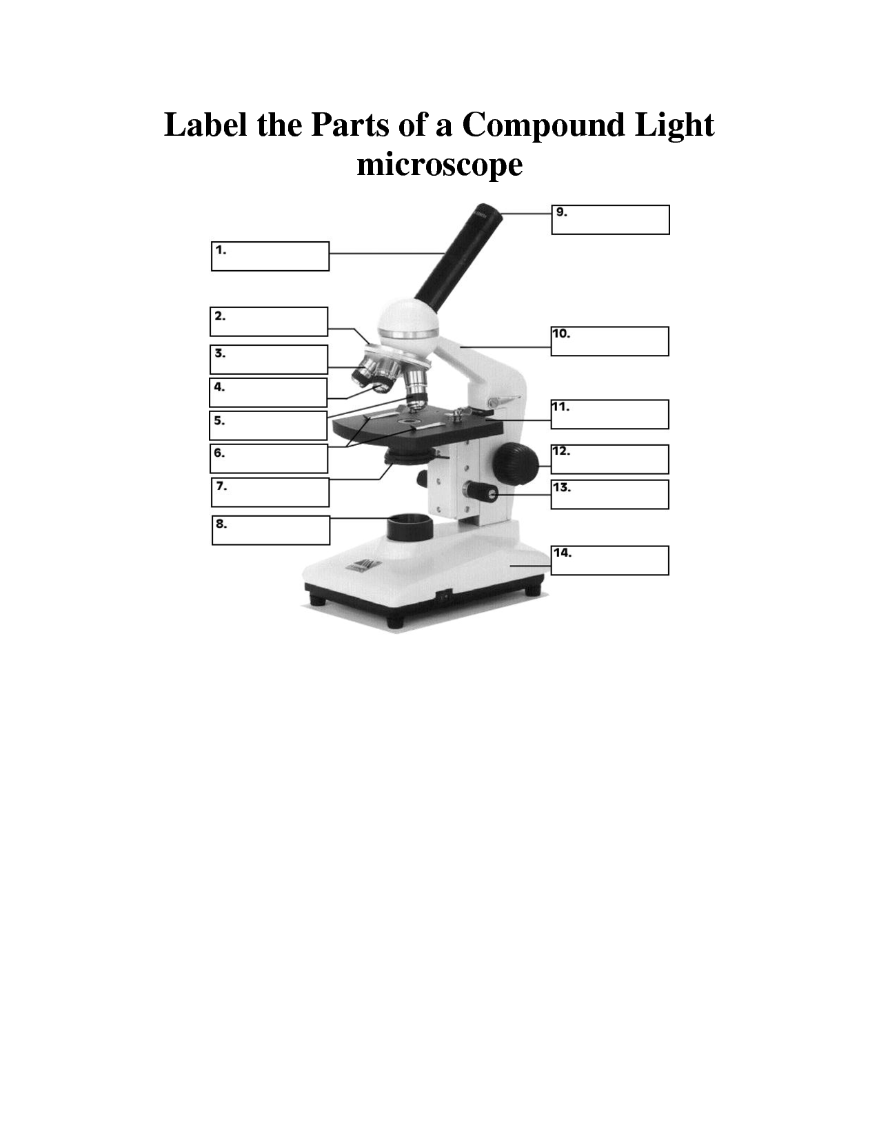
PDF Label compound microscope worksheet - Weebly [clearBoth] [clearBoth] Microscope diagram without label After you've studied all the pieces of the composite microscope, it's time to put your brain to the test. Print an unmarked microscope chart and check that you can fill out all the blanks. [clearBoth] [clearBoth] Blank microscope diagram Next we have an empty microscope diagram.

Microscope Unlabeled Diagram - Micropedia
PDF Parts of the Light Microscope - Science Spot B. NOSEPIECE microscope when carried Holds the HIGH- and LOW- power objective LENSES; can be rotated to change MAGNIFICATION. Power = 10 x 4 = 40 Power = 10 x 10 = 100 Power = 10 x 40 = 400 What happens as the power of magnification increases?
Light Microscope Diagram Labeled - Micropedia
Labeling the Parts of the Microscope | Microscope World Resources Labeling the Parts of the Microscope This activity has been designed for use in homes and schools. Each microscope layout (both blank and the version with answers) are available as PDF downloads. You can view a more in-depth review of each part of the microscope here. Download the Label the Parts of the Microscope PDF printable version here.

Female Reproductive System
Plant Cell Diagram without Labels | Plant cells worksheet ... - Pinterest Plant Cell Structure. Plant Life Cycle Worksheet. Plant Cell Project. Plant Lessons. Free plant cell worksheets for students to identify and label the parts. Younger students can use our free plant cell coloring pages, while older students can learn the parts of a cell. snoopygirl11.
8 Best Images of Lens Diagram Worksheet - Microscope with Labeled Parts, Label Eye Parts ...
Diagram of a Compound Microscope - Biology Discussion 1. It is noted first that which objective lens is in use on the microscope. 2. Stage micrometer is positioned in such a way that it is in the field of view. 3. The eyepiece is rotated so that the two scales, the eyepiece or ocular scale and the stage micrometer scale, are parallel. 4.

Best 110 Histology - Skin images on Pinterest | Anatomy, Anatomy reference and Hair follicles
A Study of the Microscope and its Functions With a Labeled Diagram Here, unlabeled microscope diagrams have been provided for your perusal, which will help you practice and test your understanding of the instrument. Types of Microscopes Depending on the source of illumination, microscopes can be divided into two categories. They are:
Biology 252 -- Plant Morphology and Systematics Home Page
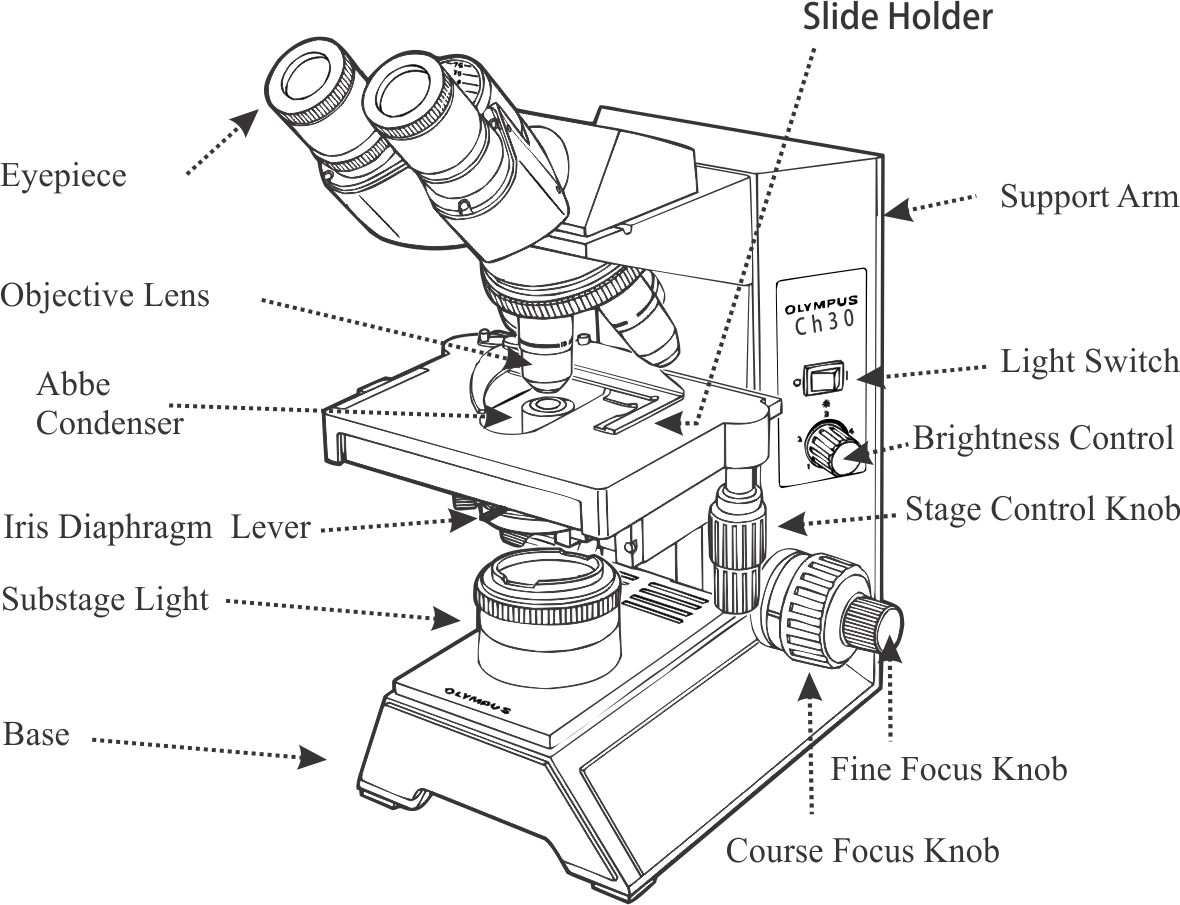
Compound Microscope Parts, Functions, and Labeled Diagram Compound Microscope Definitions for Labels. Eyepiece (ocular lens) with or without Pointer: The part that is looked through at the top of the compound microscope. Eyepieces typically have a magnification between 5x & 30x. Monocular or Binocular Head: Structural support that holds & connects the eyepieces to the objective lenses.
30 Label All Indicated Parts Of The Microscope - Labels For You
16 Parts of a Compound Microscope: Diagrams and Video Once you have an understanding of the parts of the microscope it will be much easier to navigate around and begin observing your specimen, which is the fun part! The 16 core parts of a compound microscope are: Head (Body) Arm Base Eyepiece Eyepiece tube Objective lenses Revolving Nosepiece (Turret) Rack stop Coarse adjustment knobs
Human Bio: February 2008
Animal Cell Diagram No Labels Labeled : Functions and Diagram Animal Cell Definition "An animal cell is a type of eukaryotic cell that lacks a cell wall and has a true, membrane-bound nucleus along with other cellular organelles." Explanation. Continue with more related things like blank animal cell diagram to label, labeled animal cell worksheet and. animal cell diagram without labels.
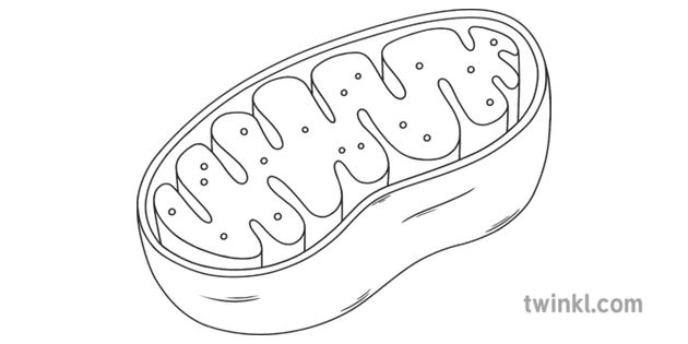
Mitochondria Science Cell Diagram Beyond Black and White RGB Illustration
Simple Microscope - Parts, Functions, Diagram and Labelling A simple microscope is a device that only has one lens for magnification. It functions the same way as the magnifying glass. Although it is simple in terms of design and function, it is useful I various fields including medicine, jewelry and watchmaking, and agriculture, to name a few. References

Microscope With Labels Clip Art at Clker.com - vector clip art online, royalty free & public domain
Junqueira's Basic Histology Text and Atlas, 14th Edition Enter the email address you signed up with and we'll email you a reset link.

the functions of a microscope | Diabetes Inc.
How Does a Microscope Work A compound microscope has two or more lenses. The eyepiece or ocular lens sits atop the body tube. Many microscopes are binocular and have two ocular lenses. Additionally, a binocular head will have a prism, either in the head or the body tube, to split the image and direct it to both oculars.

Simple Unlabelled Microscope Diagram - Micropedia
Amazing 27 Things Under The Microscope With Diagrams Amazing 27 Things Under The Microscope With Diagrams February 20, 2022 by Anupama Sapkota Note: Each image source is given below in this post of respective subheadings. Table of Contents 1. Amoeba under the microscope Direct observation Observation after staining 2. Algae under the microscope Chlorophyta Chromophyta Cryptophyta Rhodophyta

Simple Microscope Labeled Diagram - Micropedia
Post a Comment for "45 microscope diagram without labels"