39 diagram of the brain with labels and functions
Physiology, Brain - StatPearls - NCBI Bookshelf The cerebellum's primary function is to modulate motor coordination, posture, and balance. The brainstem: It contains the midbrain, pons, and medulla. It is located between the base of the cerebrum and the spinal cord. Issues of Concern Anatomical diagrams of the brain - e-Anatomy - IMAIOS These original illustrations and diagrams of the brain were created from 3D medical imaging reconstructions and then redrawn and colored using Adobe Illustrator. These anatomical charts include the main diagrams necessary for medical students, nursing students, residents, practitioners, anatomists to study the anatomy of the brain, to ...
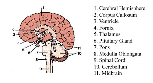
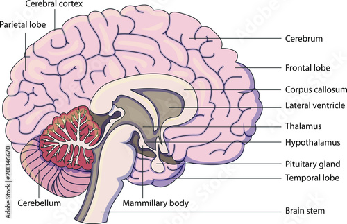
An overview of the brain - AboutKidsHealth There are three major parts of the brain: the cerebrum, cerebellum, and the brainstem. Other important areas of the brain include the thalamus, hypothalamus, pituitary gland, basal ganglia, limbic system, ventricles, cerebrospinal fluid, and 12 cranial nerves. The brain is an organ located inside your head. The brain and the spinal cord form ...

Diagram of the brain with labels and functions
Brain Ventricles: Anatomy, Function, and Conditions The human brain is fully encased in a skull to protect it from damage. For further protection, the brain is covered in three meningeal layers: pia mater, arachnoid mater, and dura mater. Pia mater is a delicate inner layer, the arachnoid mater is the middle layer consisting of a web-like structure, and the dura mater is the tough outer layer. Brain Structure and Function of Those with Bipolar Disorder Looking at a whole human brain from the outside, as shown, you see the cerebral hemispheres (the large sections, not labeled in the figure, that comprise most of the brain), the cerebellum (the small ball toward the back of the hemispheres), and the brain stem (a long, thin structure leaving the brain and connecting it to the spinal cord). The ... Circle of Willis: Anatomy, Function, and Significance Rehabilitation. The circle of Willis is a group of blood vessels in the brain that connect with each other, forming a continuous structure that resembles a circle. These nine arteries supply blood to a large portion of the brain. Most of the time, blood can flow through the vessels of the circle of Willis without any interruption.
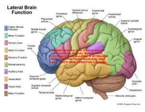
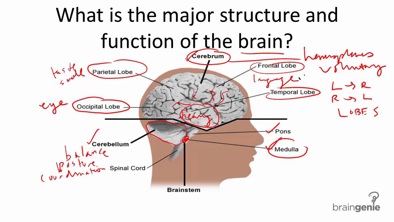
Diagram of the brain with labels and functions. What Is a Neuron? Diagrams, Types, Function, and More Takeaway. Neurons, also known as nerve cells, send and receive signals from your brain. While neurons have a lot in common with other types of cells, they're structurally and functionally unique ... What are the 12 cranial nerves? Functions and diagram Scientists use Roman numerals from I to XII to label the cranial nerves in the brain. The 12 cranial nerves include the: olfactory nerve optic nerve oculomotor nerve trochlear nerve trigeminal... Parts of the Brain Activity for Kids, Brain Diagram, and Worksheets for ... Their are 2 brain function worksheets where your student will learn about the different parts of the brain your child will learn about are: FRONTAL LOBES - The frontal lobes control voluntary movement such as reasoning, planning, parts of speech and movement, emotions, and problem-solving It is fully developed by age 10. Positions and Functions of the Four Brain Lobes | MD-Health.com The brain is divided into four sections, known as lobes (as shown in the image). The frontal lobe, occipital lobe, parietal lobe, and temporal lobe have different locations and functions that support the responses and actions of the human body. Let's start by identifying where each lobe is positioned in the brain. Position of the Lobes
Brain: Atlas of human anatomy with MRI - e-Anatomy - IMAIOS Anatomy of the brain (MRI) - cross-sectional atlas of human anatomy. The module on the anatomy of the brain based on MRI with axial slices was redesigned, having received multiple requests from users for coronal and sagittal slices. The elaboration of this new module, its labeling of more than 524 structures on 379 MRI images in three different ... Brain Ventricles: Anatomy, Function, and Conditions Your brain's ventricular system is comprised of four ventricles as well as small structures that connect each ventricle called foramina. The first and second ventricles are lateral ventricles. These C-shaped structures are located on each side of your cerebral cortex, the wrinkly outer layer of your brain. Structures Of The Brain Quiz! Ultimate Trivia - ProProfs Questions and Answers 1. This structure controls thought, voluntary movement, language, reasoning and perception. A. Cerebellum B. Cerebral cortex C. Cerebrum 2. This is a collection of axons that connect the right and left hemispheres of the brain. A. Corpus callosum B. Cerebral hemisphere C. Corporal callosum 3. Brain: Ultimate Guide to the Brain for AP® Psychology - Albert The forebrain consists of the thalamus, hypothalamus, amygdala, and the hippocampus. The hypothalamus, amygdala, and hippocampus make up what we call the Limbic System of your brain. Thalamus The thalamus is located between the cerebral cortex and the midbrain. It is made up of nuclei that receive different sensory and motor inputs.
Parts of the brain: Learn with diagrams and quizzes | Kenhub Labeled brain diagram First up, have a look at the labeled brain structures on the image below. Try to memorize the name and location of each structure, then proceed to test yourself with the blank brain diagram provided below. Labeled diagram showing the main parts of the brain Blank brain diagram (free download!) Structure of the Brain and Their Functions - New Health Advisor The function of these lobes is listed below: Frontal lobe- Associated with planning of speech, reasoning, emotions, problem solving and movement. Occipital lobe- It's associated with visual processing Parietal lobe- It's associated with recognition, movement, orientation, perception of stimuli, speech and memory. Left Brain vs. Right Brain: Characteristics Chart [INFOGRAPHIC] Your right hemisphere tries to call out the color, while your left side of the brain focuses on the words' meanings. If you practice a little, this funny task will become much more manageable. Practice makes perfect for every skill! That's why we have collected some useful exercises that will help you develop both your left and right brain. Left and Right Hemisphere of the Brain | Functions & Characteristics The left hemisphere of the brain is in-charge of the cognitive functions such as speech and language. The right hemisphere of the brain is more on creativity and face recognition. Although the functions of the brain is divided based on its hemisphere, even a particular functions to be executed, it would still need the entire brain.
Addiction and the Brain: Areas of the Brain Affected by Addiction | Willow Place for Women
Parts Of The Brain Quiz Questions And Answers - ProProfs Questions and Answers 1. What is the biggest part of the brain? A. Cerebrum B. Thalamus C. Brain stem D. Cerebellum 2. What is the back part of the brain called? A. Thalamus B. Cerebellum C. Cerebrum D. Brain stem 3. What sits below the cerebrum and in front of the cerebellum? A. Cerebellum B. Brainstem C. Thalamus D. Cerebrum 4.
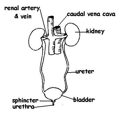
Excretory System Answers - WikiEducator
Brain: Function and Anatomy, Conditions, and Health Tips Some of its main functions include: processing sensory information regulating blood pressure and breathing releasing hormones Brain diagram Use this interactive 3-D diagram to explore the brain....
Human Brain Illustrations | MedicalNDX
14 Informative Facts, Diagram & Parts Of Human Brain For Kids The cerebrum, divided into four 'lobes' or regions, controls higher functions, such as learning, reasoning and speech, and senses, such as vision and hearing. Occipital lobe: Located at the back of the cerebrum, the occipital lobe is the primary visual area of the brain. The occipital lobe processes visual information for your eyes.
(209).jpg)
Quiz: Physiology Of The Brain And Its Various Functions - ProProfs Quiz
Human Brain Lesson for Kids: Function & Diagram - Study.com Your cerebrum also controls how you speak, learn, remember things, and feel emotions, like happiness and sadness. It even helps you understand what's going on around you by receiving messages from...
Label Diagram Of The Brain - Wiring Diagram
Brain - Wikipedia A brain is an organ that serves as the center of the nervous system in all vertebrate and most invertebrate animals. It is located in the head, usually close to the sensory organs for senses such as vision.It is the most complex organ in a vertebrate's body. In a human, the cerebral cortex contains approximately 14-16 billion neurons, and the estimated number of neurons in the cerebellum is ...

Solved: Label the structures of the brain indicated in the foll... | Chegg.com
Parts of the Human Brain | Anatomy & Function - Study.com The parts of the brain include the cerebrum, the cerebellum, the brain stem, and the pituitary gland. The brain structure is protected by the skull, which is composed of the cranium and the bones...

Critical Literacy.. :): Brain and it's function...
Brain Structures and Their Functions | MD-Health.com It is divided into four sections: the temporal lobe, the occipital lobe, parietal lobe and frontal lobe. The cerebrum is divided into a right and left hemisphere which are connected by axons that relay messages from one to the other.

The parts of the brain and the functions. Labeled diagram. | The Brain | Pinterest ...
Anatomy of the Brain - Simply Psychology The temporal lobes are located on both sides of the brain, near the temples of the head, hence the name temporal lobes (Figure 5). The main functions of these lobes include understanding, language, memory acquisition, face recognition, object recognition, perception, and processing auditory information.
Brain diagram labeled
Human brain - Wikipedia The main functions of the frontal lobe are to control attention, abstract thinking, behaviour, problem solving tasks, and physical reactions and personality. [24] [25] The occipital lobe is the smallest lobe; its main functions are visual reception, visual-spatial processing, movement, and colour recognition.
8 1 1 Brain Structure and Function - YouTube
Lobes of the brain: Structure and function | Kenhub The lobes of the cerebrum are actually divisions of the cerebral cortex based on the locations of the major gyri and sulci. The cerebral cortex is divided into six lobes: the frontal, temporal, parietal, occipital , insular and limbic lobes. Each lobe of the cerebrum exhibits characteristic surface features that each have their own functions.
Label Diagram Of The Brain - Drivenheisenberg
Circle of Willis: Anatomy, Function, and Significance Rehabilitation. The circle of Willis is a group of blood vessels in the brain that connect with each other, forming a continuous structure that resembles a circle. These nine arteries supply blood to a large portion of the brain. Most of the time, blood can flow through the vessels of the circle of Willis without any interruption.

Anatomy and Functions of the Brain Medical Illustration
Brain Structure and Function of Those with Bipolar Disorder Looking at a whole human brain from the outside, as shown, you see the cerebral hemispheres (the large sections, not labeled in the figure, that comprise most of the brain), the cerebellum (the small ball toward the back of the hemispheres), and the brain stem (a long, thin structure leaving the brain and connecting it to the spinal cord). The ...

Labeled Parts Of The Brain - koibana.info | Brain diagram, Brain anatomy and function, Brain anatomy
Brain Ventricles: Anatomy, Function, and Conditions The human brain is fully encased in a skull to protect it from damage. For further protection, the brain is covered in three meningeal layers: pia mater, arachnoid mater, and dura mater. Pia mater is a delicate inner layer, the arachnoid mater is the middle layer consisting of a web-like structure, and the dura mater is the tough outer layer.

Brain anatomy and function - lateral and sagittal view — Medical Art Works

What do the different parts of your brain control... well, here you go! | Mental Health ...

Brain Function Diagram Stock Vector 156466463 - Shutterstock

Psycholinguistics : Language And The Brain ~ My Inspiration
Post a Comment for "39 diagram of the brain with labels and functions"